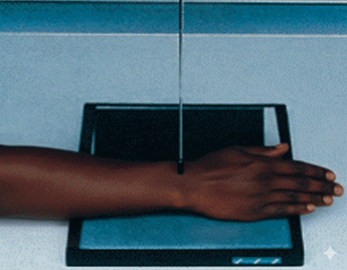
AP OBLIQUE WRIST PROJECTION
Anteroposterior oblique projection for evaluation of the medial carpus, pisiform, and hamate

Exposure Factors
Optimal parameters: Similar to standard wrist projection but with specific rotation.
Visible Anatomical Structures
This projection specifically highlights the medial column of the carpus:
- Pisiform: Observed in profile, separated from the triquetrum.
- Hamate (Hook of Hamate): Better visualization of its medial aspect.
- Triquetrum: Clearer view of its joint surfaces.
- Distal Radio-ulnar Joint: Evaluated from an oblique perspective.
- Ulnar Styloid Process: Clearly visible.
Image Receptor Size and Orientation
One half of the plate can be used if comparing with another projection.
Patient Positioning
Central Ray Specifications
Point of Entry: Mid-carpal area (midpoint between the styloid processes).
Direction: Straight to the center of the image receptor.
Collimation: Must include distal radius/ulna and proximal metacarpals.
Acceptable Image Criteria
Rotation
Medial 45° rotation clearly evident.
Separation
Pisiform seen in profile and separated.
Detail
Clear trabecular patterns of carpal bones.
Overlap
Minimum superimposition of the distal ulna.
Clinical Selection: AP vs PA Oblique
AP Oblique (Medial Rotation) is indicated for:
- Medial carpal pain (Pisiform, Triquetrum, Hamate).
- Evaluation of the Pisiform without superimposition.
- Ulnar side trauma of the wrist.
- Foreign bodies in the medial area.
PA Oblique (Lateral Rotation) is indicated for:
- Lateral carpal pain or trauma.
- Suspected Scaphoid or Trapezium fracture.
- Osteoarthritis evaluation of lateral joints.
- Rhizarthrosis (Trapeziometacarpal arthritis).
Choose based on the location of symptoms or the structures to be evaluated.